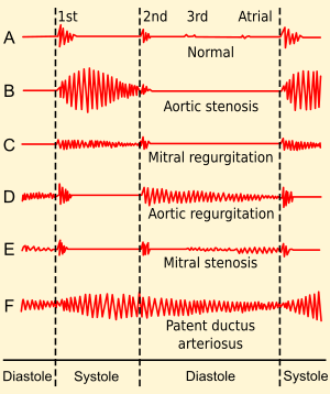
heart sounds
During each cardiac cycle two prominent sounds are produced which can be easily heard through a stethoscope.
First Heart Sound (LUBB) | Second Heart Sound (DUB) |
1. It is produced by closing of AV valves (tricuspid valves) during ventricular systole. | 1. It is produced by closing of semilunar valves at the beginning of ventricular diastole. |
2. It is low pitched and of longer duration. | 2. It is higher pitched and of shorter duration. |
REGULATION OF CARDIAC ACTIVITY:-
Normal activities of the heart are regulated intrinsically i.e autoregulated by specialized muscles (nodal tissue), hence the heart is called myogenic. This automatic rhythm is produced by the spontaneous depolarization, which leads to contraction of heart.
A special neural centre in the medulla oblongata (in the brain) can moderate the cardiac function through autonomic nervous system (ANS). Sympathetic and parasympathetic nerves (parts of ANS) are connected to the heart and can modify the rate of spontaneous depolarization of the SA node.
Sympathetic nerve endings release noradrenaline which stimulates the SAN that accelerates the heart beat, the strength of ventricular contraction and thereby the cardiac output. On the other hand, parasympathetic nerve endings release acetylcholine which decreases the rate of the heart beat, speed of conduction of action potential and thereby the cardiac output.
The adrenal medulla secretes two hormones called adrenaline and noradrenaline. Both the hormones are rapidly secretes in response to stress of any kind and during emergency situations. These hormones increase the heart beat and the strength of heart contraction. Thus, adrenal medullary hormones can also increase the cardiac output.
Note:-
- High level of potassium and sodium ions decrease heart rate and strength on contraction.
- Ab excess of calcium ions increases heart rate.
- Increased body temperature during fever increases heart rate.
- Strong emotions such as fear, anger and anxiety increase heart rate, resulting in increased blood pressure.
ELECTROCARDIOGRAM (ECG)
The pacemaker region of the heart (SA node) exhibits a spontaneous depolarization that causes action potentials, resulting in the automatic beating of the heart. Impulses which travel through cardiac muscles during the cardiac cycle produce electrical currents. The electrical currents are conducted through the body fluids to the body durface, where the amplified currents can be detected by placing electrodes on the skin and recorded as an electrocardiogram (ECG). It was discovered by Einthoven.
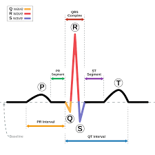
Electrocardiogram (ECG), is a graphical representation of the electrical activity of the heart during a cardiac cycle. The machine used to obtain an electrocardiogram is known as electrocardiograph and this technique is called electrocardiography.
To obtain a standard ECG, a patient is connected to the machine with three electrical leads (one to each wrist and one to the left ankle).
- The P wave is a small upward wave that represents electrical excitation (or depolarisation) of the atria which leads to contraction of both the atria.
- The QRS (wave) complex represents the depolarization of the ventricles, which initiates the ventricular contraction (ventricular systole). The contraction of the ventricles starts shortly after Q and marks the beginning of the systole.
- The T-wave represents the return of the ventricles from excited (depolarised) to normal state (e repolarisation). The end of the T-wave marks the end of systole.
- Thus, by counting the number of QRS complexes that occur in a given time period, the heart beat rate of an individual can be determined. ECG is of great clinical significance as any deviation from this shape indicates a possible abnormality or disease.
- Enlargement of P-wave indicates enlargement of the atria.
- The enlarged Q and R waves indicates myocardial infarction (heart attack).
- The S-T segment is elevated in myocardial infarction and depressed when the heart muscles receive insufficient oxygen.
- T-wave is flat when the heart muscles receive insufficient oxygen as in atherosclerotic heart disease.
The monitoring machine (electro- cardiograph) makes the sound “…pip …pip …pip…. Peeeeeeeeeeeeeeeee” as the patient goes into cardiac arrest
Note:-
- Artificial pacemaker:- The SA node is the natural pacemaker of heart. When it does not work properly, an artificial pacemaker is implanted so that the normal heart beat can be restored and maintained. The artificial pacemaker was introduced by Chardack in 1960.
- In patients exhibiting symptoms of ventricular escape (Stokes-Adam- Syndrome) in which the atrial impulse suddenly fails to be transmitted to the ventricle, the artificial pacemaker is connect to the right ventricle for controlling its rhythm.
Body fluids include blood, lymph, cerebrospinal fluid, interstitial fluid, and intracellular fluid. They are essential for transporting nutrients, oxygen, and waste products throughout the body.
The circulatory system is a network of organs and vessels, including the heart, blood, and blood vessels, responsible for transporting blood, nutrients, hormones, oxygen, and other gases to and from cells.
The heart functions as a pump with four chambers: two atria and two ventricles. The right side pumps deoxygenated blood to the lungs, while the left side pumps oxygenated blood to the rest of the body.
Blood consists of plasma (the liquid part), red blood cells (carry oxygen), white blood cells (fight infection), and platelets (help with clotting).
Plasma, which makes up about 55% of blood volume, is a yellowish fluid that carries nutrients, hormones, and proteins throughout the body. It also helps maintain blood pressure and volume.
There are two main types of blood circulation: systemic circulation (throughout the body) and pulmonary circulation (through the lungs).
The lymphatic system is a network of tissues and organs that help rid the body of toxins and waste. It includes lymph nodes, lymphatic vessels, and lymph, which is a fluid containing infection-fighting white blood cells.
Red blood cells contain hemoglobin, a protein that binds to oxygen in the lungs and releases it to tissues throughout the body.
High blood pressure, or hypertension, can be caused by factors such as genetics, poor diet, lack of exercise, obesity, stress, and chronic conditions like diabetes.
Common disorders include hypertension, atherosclerosis (hardening of the arteries), heart attack, stroke, and aneurysms.