Introduction:-
Proteins are large-sized macromolecules having one or more polypeptides (chains or polymers of amino acids linked by peptide bond).
As there are 20 types of amino acids, a protein is a heteropolymer and not a homopolymer. A homopolymer has only one type of monomer repeating ‘n’ number of times
Collagen is the most abundant protein in animal world. It is the main component of connective tissue of animals.
Ribulose bisphosphate carboxylase-oxygense (RubisCo) is the most abundant protein in the whole of the biosphere
structure of proteins:-
Biologists describe the protein structure at four levels-
(i) primary structure:- The sequence in which amino acids are arranged in a polypeptide chain of a protein is called its primary structure. It gives the positional information of amino acids in a protein i.e.; which is the first amino acid which is the second and so on.
In this, chains of amino acids which constitute a protein, the amino acid present at the left end is the first amino acid, whereas the one at the right end is the last amino acid of the protein. The first amino acid is known as N-terminal amino acid and the last is known as C-terminal amino acid.
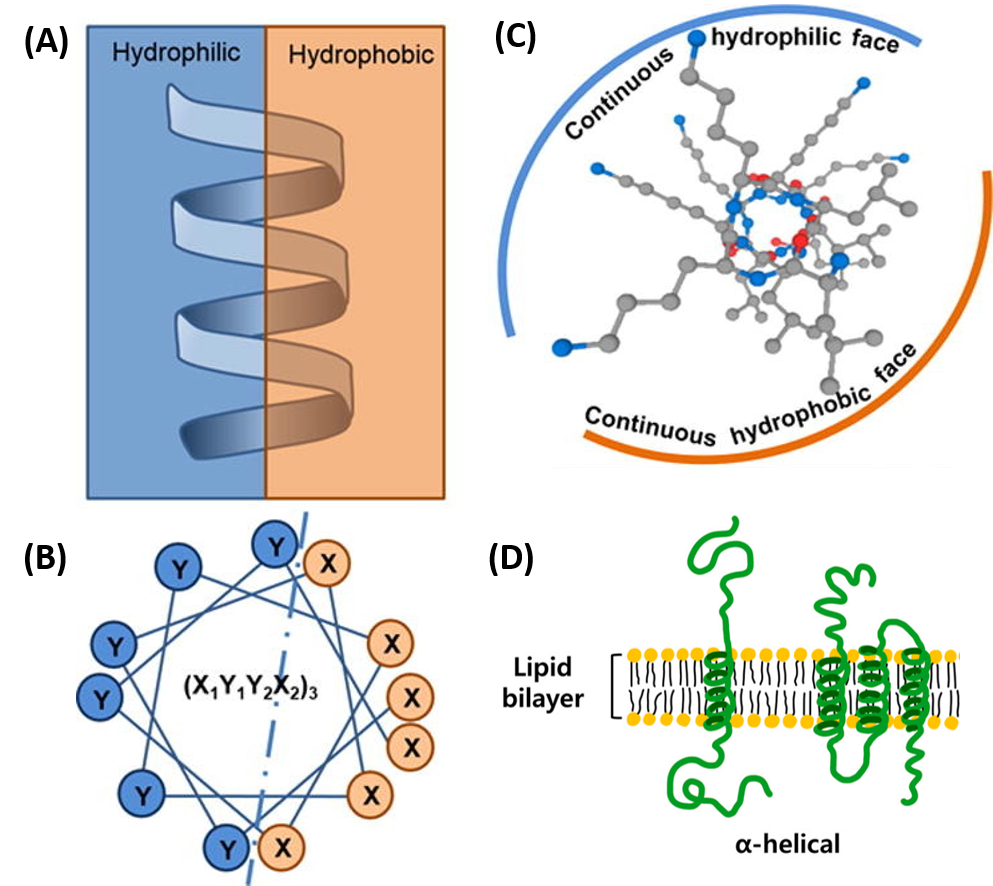
(ii) Secondary structure:- Some protein of the protein thread are folded either in the form of a helix ( similar to a revolving staircase) Betta-pleated sheet. The Alpha-helix and Betta-pleated sheet are two types of secondary structures. In Alpha-hilix, there is interaction between every fourth amino acid by the formation of intramolecular hydrogen bond.
The polypeptide gets a helical shape (Alpha-helix). The intramolecular hydrogen bond is a bond formed between hydrogen atom and the highly electronegative atom such as nitrogen, oxygen and fluorine of the same molecule.
In proteins, only right-handed belices are observed.
Example:- Keratin protein present in hair.
Collagen helix:- The polypeptide coil of collagen helix is strengthened by establishing hydrogen bond and locking effect also occurs with the help of amino acids proline and hydroxyproline.
(iii) Tertiary structure:- The long protein chain or the polypeptide chain usually folds upon itself like a hollow woolen ball. This is termed as the tertiary structure. This structure gives a 3-dimensional view of a protein
Tertiary structure is absolutely necessary for the many biological activities of proteins for example, this structure brings distant amino acid side chains closer forming the active site (the site to which a substrate gets attached) of proteins i.e.; enzymes.
Example:- Myoglobin (protein found in muscle cells)
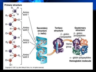
(iv) Quaternary structure:- Quaternary structure is formed when a protein has more than one subunits (individual polypeptide chains of a quaternary protein are called subunits) or polypeptide chains and each polypeptide has a primary, secondary or tertiary structure of its own.
The way in which these individually folded polypeptides are arranged with respect to each other gives the architecture of the quaternary structure of a protein.
Example:- Haemoglobin has such structure. Haemoglobin has four helical polypeptide chains, two Alpha-chains and two Betta-chains.
Functions:-
Enzymatic Catalysis: Enzymes, which are proteins, accelerate biochemical reactions. They are essential for metabolism, DNA replication, and many other cellular processes
Structural Support: Proteins like collagen, elastin, and keratin provide structural support to cells and tissues. Collagen, for example, is a major component of connective tissues.
Transport and Storage: Hemoglobin, a protein in red blood cells, transports oxygen from the lungs to tissues throughout the body. Other proteins transport ions, molecules, and nutrients across cell membranes or within cells
Immune Response: Antibodies (immunoglobulins) are proteins that recognize and bind to specific antigens, such as bacteria and viruses, to neutralize them and help protect the body from infection.
Cell Signaling: Proteins such as receptors and hormones are involved in signaling pathways that regulate physiological processes. Insulin, for example, is a hormone that helps regulate blood glucose levels
Movement: Motor proteins like myosin and actin are involved in muscle contraction and movement. They convert chemical energy into mechanical work, which is essential for locomotion and other cellular movements.
Regulation of Gene Expression: Proteins such as transcription factors bind to specific DNA sequences and regulate the transcription of genes, thus controlling the levels and timing of gene expression.
Cellular Communication: Membrane proteins act as receptors for signaling molecules, facilitating communication between cells and their external environment. They are involved in various signaling pathways that control cell growth, differentiation, and apoptosis.
Storage Proteins: Some proteins serve as storage forms of amino acids, which can be utilized when needed. Ferritin, for example, stores iron in the liver.
Defensive Proteins: In addition to antibodies, proteins like complement proteins assist in the defense against pathogens by enhancing the ability of antibodies and phagocytic cells to clear microbes and damaged cells from an organism.
Hormonal Proteins: Hormonal proteins act as messengers, coordinating various physiological activities. Examples include insulin, growth hormone, and thyroid hormones.
Conjugated proteins are formed by the binding of a simple protein with a non- protein part called the prosthetic Group.
The conjugated proteins are of the following types:-
(a) Nucleoproteins (prosthetic group-nuclei acid) e.g.; protamines
(b) Metalloproteins (prosthetic group-metals) e.g.; Ferritin
.
(c) Chromoproteins (prosthetic group-pigment) e.g; Cytochromes
(d) Phosphoproteins (prosthetic group-phosphoric acid) e.g.; Casein of milk.
(e) Lipoproteins (prosthetic group-lipids) e.g.; Chylomicron.
(f) Glycoproteins (prosthetic group-carbohydrates) e.g.; Mucin.
The primary structure, or amino acid sequence, determines the protein’s unique characteristics and dictates how it will fold into its specific three-dimensional shape, which is crucial for its function. Even a single change in the amino acid sequence can significantly impact the protein’s structure and function.
Alpha-helices form when the polypeptide chain coils into a spiral shape stabilized by hydrogen bonds between the carbonyl oxygen of one amino acid and the amide hydrogen of another, typically four residues apart. Beta-sheets form when segments of the polypeptide chain align side-by-side, either in parallel or antiparallel orientation, with hydrogen bonds forming between the carbonyl oxygen of one strand and the amide hydrogen of an adjacent strand.
Disulfide bridges (or disulfide bonds) are covalent bonds that form between the sulfur atoms of two cysteine residues within or between polypeptide chains. These bonds provide significant stability to the protein’s tertiary and quaternary structures, helping to maintain its functional conformation, especially in extracellular environments.
Protein structure can be determined using various techniques, including:
X-ray Crystallography: This method involves crystallizing the protein and analyzing the diffraction patterns of X-rays passing through the crystal to determine the atomic structure.
Nuclear Magnetic Resonance (NMR) Spectroscopy: This technique uses the magnetic properties of atomic nuclei to determine the three-dimensional structure of proteins in solution.
Cryo-Electron Microscopy (Cryo-EM): This technique involves rapidly freezing the protein and capturing images with an electron microscope to reconstruct the protein’s structure at near-atomic resolution.