Introduction:-
Urine formation is a charismatic and complicated process that exemplifies the body’s remarkable ability to maintain internal balance and homeostasis
This vital biological function begins in the kidneys, where an elaborate system of filtration, reabsorption, and secretion meticulously orchestrates the removal of waste products and excess substances from the bloodstream.
It involves three main processes namely, glomerular filtration, reabsorption and tubular secretion, that takes place in different parts of the nephron.
Glomerular Filtration:-
The first step in urine formation is the filtration of blood, which is carried out by the glomerulus and is called glomerular filtration. On an average 1100-1200 ml of blood is filtered by the kidneys per minute which constitute roughly 1/5th of the blood pumped out by each ventricle of the heart in minute.
The glomerular capillary blood pressure causes filtration of blood through three layers i.e
(i) The endothelium of glomerular blood vessels
(ii) The epithelium of Bowman’s capsule
(iii) A basement membrane between these two layers
These three structures form the filtration membrane.
The epithelial cells of Bowman’s capsule called podocytes are arranged in an intricate manner so as to leave some minute spaces called filtration slits or slit pores.
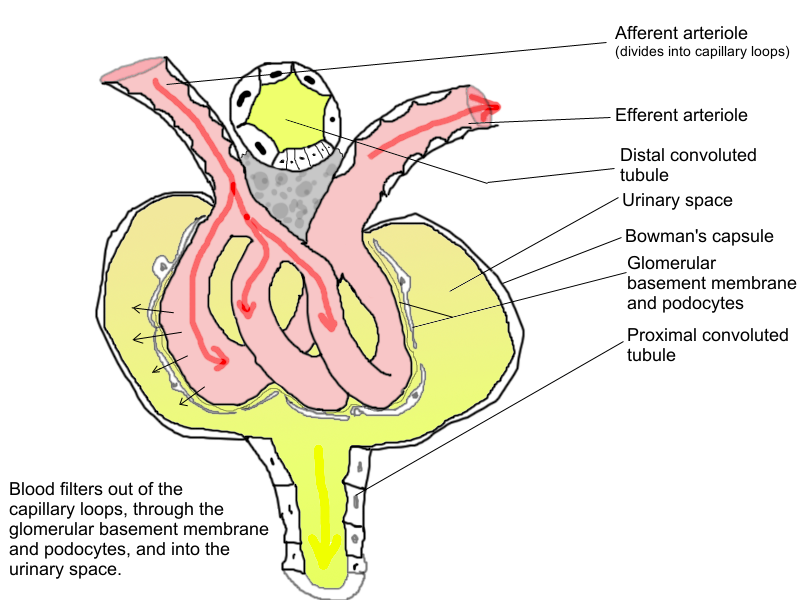
Blood filtered so finely through these membranes, that almost all the constituents of the plasma except the proteins pass into the lumen of the Bowman’s capsule.Therefore, it is considered as a process of ultrafiltration.
Development of filtration pressure:- Blood flows through glomerular capillaries under a pressure. This pressure is due to two reasons:
Larger diameter of afferent arteriole as compared to smaller diameter of efferent arteriole.
Natural arterial pressure is caused by pumping activity of heart. Blood pressure 60 mm Hg, this is called glomerular hydrostatic pressure (GHP). Osmotic concentration of proteinaceous content of glomerular blood is equivalent to 30mm hg, this is called blood colloidal osmotic pressure (BCOP).
Pressure of interstitial fluid and pressure of renal filtrate is collectively called capsular hydrostatic pressure (CHP= mm Hg). The pressure being extended on glomerular blood for undergoing filtration is called glomerular filtration pressure (GFP). It is about 10-25 mmHg.
or,
GFP = GHP – (BCOP + CHP)
= 60 – (30+20)
= 10 mm Hg
The amount filtrate formed by the kidneys per minute is calle glomerular filtration rate (GFR). GFR in a healthy individual is approximately 125 ml/min, i.e., 180 litters per days.
About 700 ml of plasma flows per minute through the kidney (RPF, renal plasma flow). Out of it, about 125 ml of filtrate is formed by glomeruli per minute. Therefore, the normal filtration fractionis GFR/RPF i.e 125/700 = 0.18 or 18%.
Autoregulation of glomerular filtration
There are three methods by which renal blood flow and glomerular filtration rate (GFR) are automatically regulated
(i) Myogenic autoregulation:- A rise in blood pressure should normally increase blood flow through glomeruli. However, stretching
of the vascular wall inceases passage of Ca2+ ions from extracellular fluid into the cells resulting in their contraction. Contraction checks overstretching of vascular walls and raises vascular resistance so that rate of blood flow and GFR are brought down to normal.
(II) Juxtaglomerular apparatus (JGA) :- JGA is a special sensitive region formed by cellular modifications in the distal convoluted tubule and the afferent at the location of their contact. The JGA becomes active when there is decrease in renal blood pressure or decrease in GFR. It promotes release of rennin from juxtaglomerular cells
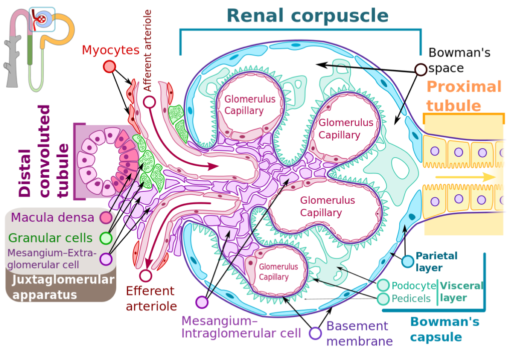
This rennin converts protein angiotensinogen into peptide angiotensin. Angiotensin is a hormone that raises glomerular blood pressure through constricting efferent arteriole resulting in restoring GFR.
(iii) Neural control:- Blood vessels of the kidney are innervated by nerve fibres of the sympathetic nervous system. When activated, the nerve fibres bring about constriction of renal arteries and cause decrease in renal blood flow as well as glomerular filtration rate..
Tubular Reabsorption:-
A comparison of the volume of the filtrate formed per day (180 litres per day) with that of the urine released (1.5 litres), suggested that nearly 99 percent of the filtrate has to be reabsorbed by the renal tubules.
This process is called reabsorption. The tubular epithelial cells in different segments of nephron perform this either by active or passive mechanisms. For example, substances like glucose, aminoacids, Na+ , etc.,in thefiltrate are reabsorbed activity, whereas the nitrogenous wastes are absorbed by passive transport. Reabsorption of water also occurs passively in the intial segments of the nephron.
Tubular Secretion
The phenomenon of secretion of metabolic wastes by tubular cells into the filtrate is known as tubular secretion. It includes secretion of K+ , H+ , ammonia and some wastes like hippuric acid, creatinine, drugs, pigments, toxins etc.
It is the only mode of excretion in animals which do not have glomeruli, e.g , desert amphibians, marine fishes. Tubular secretion is also an important
step in urine formation as it helps in the maintenance of ionic and acid-base balance of body fluid.
The primary function of the kidneys in urine formation is to filter waste products and excess substances from the blood to form urine. This process helps regulate the body’s fluid balance, electrolyte levels, and the removal of toxins and metabolic waste products.
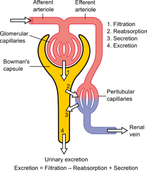
Urine formation involves three main stages:
Filtration: Blood is filtered in the glomerulus of the nephron, where water, ions, and small molecules pass into the renal tubule, forming the filtrate.
Reabsorption: Essential substances like glucose, amino acids, and water are reabsorbed from the filtrate back into the bloodstream in the proximal convoluted tubule, loop of Henle, distal convoluted tubule, and collecting duct.
Secretion: Additional waste products and excess ions are secreted from the blood into the renal tubule to be included in the urine.